Description
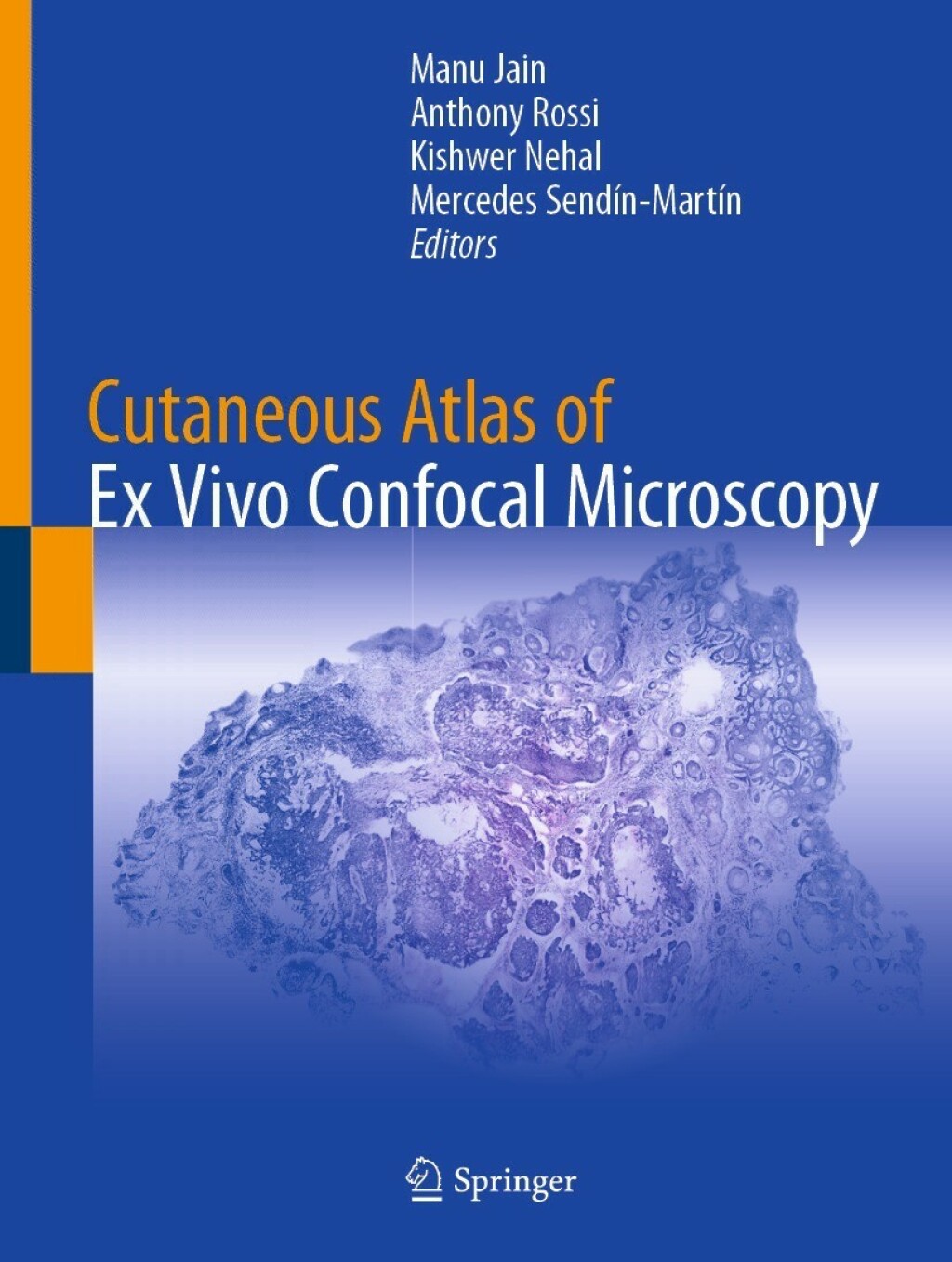
This atlas provides a detailed overview of the novel technique of ex vivo confocal microscopy for rapid imaging of excised tissues in dermatological practice. It features an extensive collection of ex vivo images�acquired from normal skin structures and from a variety of neoplastic lesions (benign and malignant) and inflammatory lesions. Each chapter contains several image types of a particular disorder, including�gray-scale, digital purple-pink images�(DHE) and�hematoxylin and eosin (H&E) correlations to assist the acquisition of diagnostic skills. Guidance on how to use techniques for tissue preparation, staining, handling and�image acquisition are also provided enabling the reader to develop confidence in integrating this technique into their day-to-day practices. Furthermore, this atlas also provides an update on the ongoing latest advances in the field. � Cutaneous Atlas of Ex Vivo Confocal Microscopy�covers how to apply these techniques into dermatological practice, especially in Mohs surgery for the evaluation of keratinocytic neoplasm and in dermatopathology for rapid evaluation of�varied skin lesions. It is therefore a valuable resource for trainee, residents, practicing dermatologists and dermatopathologists who are seeking a resource to assist in developing their knowledge and skills of utilizing these methodologies.�Typham this is the title: Cutaneous Atlas of Ex Vivo Confocal Microscopy


Reviews
There are no reviews yet.